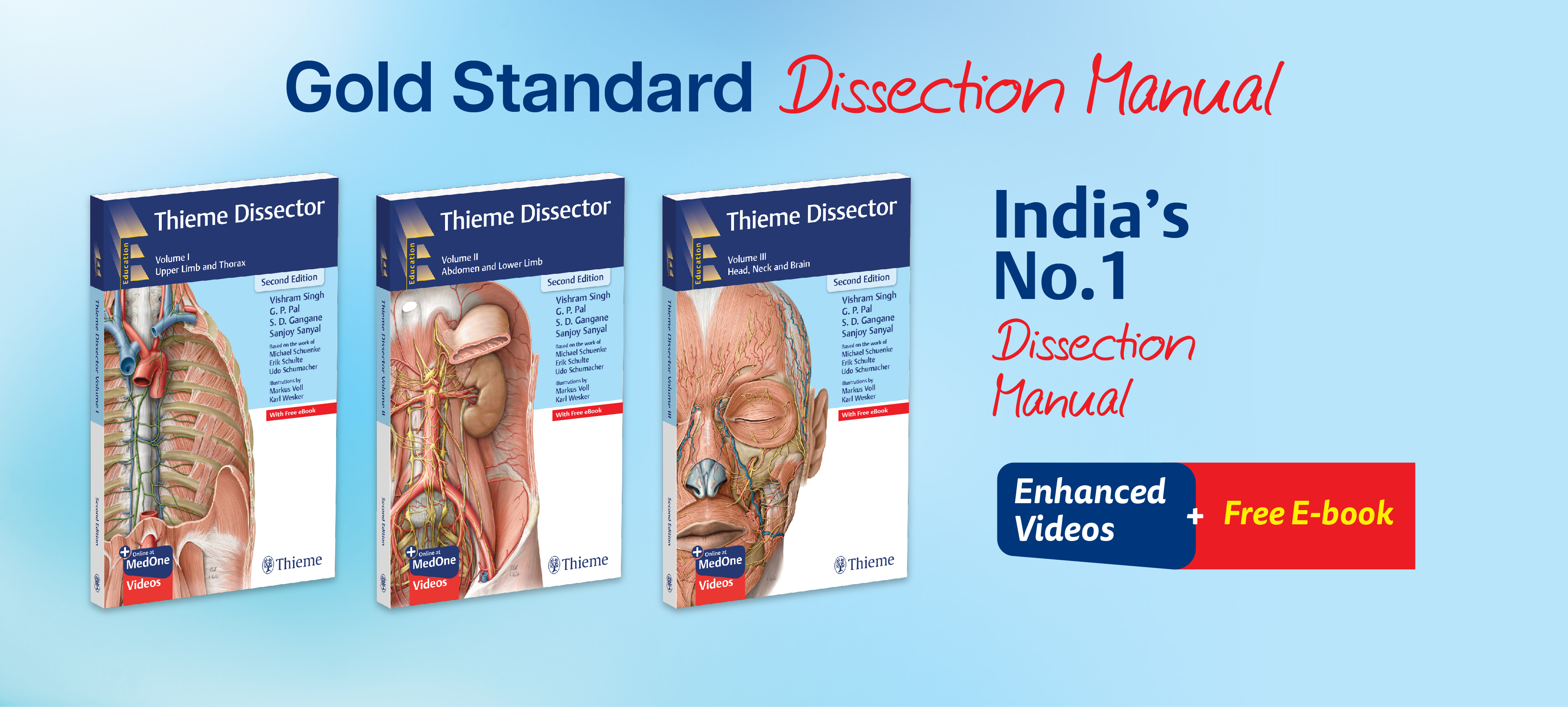
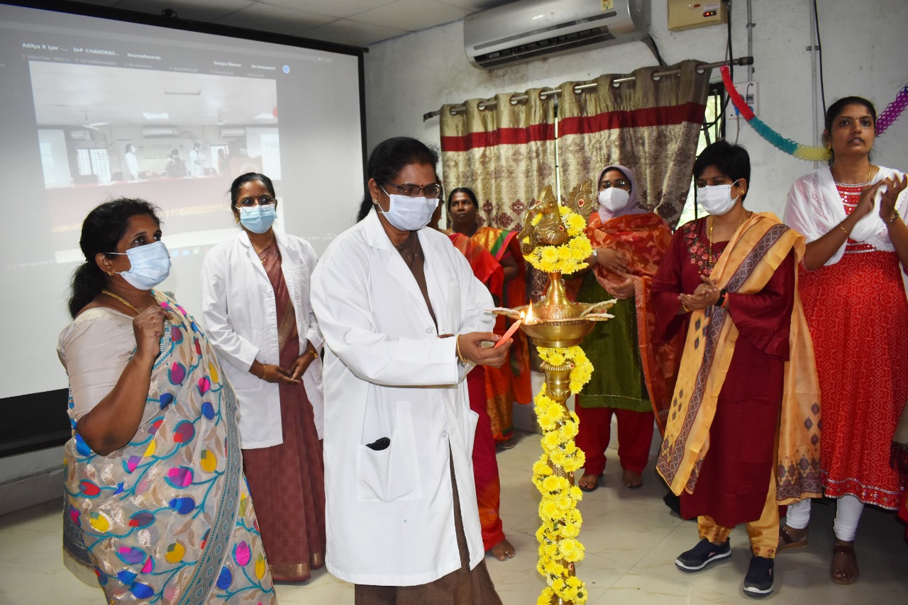
About the book
Thieme Dissector, Second Edition is a richly illustrated and detailed three-volume manual for guiding students and teachers in the dissection lab. It is enriched with intricate illustrations of the human anatomy created from the clinician’s perspective. These illustrations and the carefully researched and structured text elucidate the layer-by-layer dissection of each region of the human body in a stepwise manner. The volumes have been created by renowned experts in the field: Dr. Vishram Singh, Dr. G. P. Pal, Dr. S. D. Gangane, and Dr. Sanjoy Sanyal. The text of the volumes flows lucidly through well-defined sections in each chapter. These sections have also been made visually distinct to aid access. The authors have aimed to make the reading of these volumes educative, interesting, and visually engaging.
Salient Features of the Second Edition
- Updated videos: Provides access to more than 100 new videos on Thieme MedOne to facilitate learning, understanding, and comprehension. These videos enhance the scope of understanding the topic under discussion.
- Dissection screenshots: Most relevant and duly labelled screenshots from the cadaveric dissection videos are presented at suitable places within the text to provide better insight into the steps of dissection.
- Radiographs: Includes newly added radiographs to help broaden the gamut of interpretation of the anatomy.
- New section: A new section on “Vertebral Column” has been added to Volume I for extensive coverage of the back region.
Features
-
Best in ContentThe set of three volumes have been put together by the most eminent medical practitioners and celebrated authors in this field. These distinguished authors have meticulously and thoroughly explained anatomical relations and concepts
in lucid language, with the help of excellent illustrations. The text is divided in easy-to- understand and remember sections. These sections have also been made visually distinct for further ease of readers. The illustrations have detailed labelling and captions.
-
Lifelike IllustrationsThe three volumes include over 600 original, realistic illustrations created by the best medical illustrators. These illustrations help present anatomical information in detail, with unprecedented clarity. They form the perfect companion to the information provided in the text.
-
Dissection VideosThe three volumes are accompanied by 200 dissection videos that will help students get a near-real experience of being a part of the dissection process.

-
AuthorsDr. Vishram Singh MBBS, MS, PhD (hc), MICPS, FASI, FIMSA, Former Professor and Head, Department of Anatomy, Santosh Medical College; Member of Academic Council and Core Committee PhD Course, Santosh University, Ghaziabad, Delhi NCR, India; Editor-in-Chief, Journal of the Anatomical Society of India.Dr. G.P. Pal MBBS, MS, DSc, FASI, FAMS, FNASc, FASc, Bhatnagar Laureate, Director Professor, Index Medical College, Indore, Madhya Pradesh, India.Dr. S. D. Gangane MBBS, MS, FAIMS, Professor and Head, Department of Anatomy, Terna Medical College, Navi Mumbai, Maharashtra, India.Dr. Sanjoy Sanyal MBBS, MS (Surgery), MSc (Royal College of Surgeons of Edinburgh), ADPHA, Provost and Dean of Academic Affairs, Professor and Department Chair of Anatomical Sciences, Richmond Gabriel University College of Medicine, St. Vincent and the Grenadines, Surrey, Canada.


Send Message to Authors!
Quintessential Atlas of Anatomy expands on widely acclaimed prior editions!
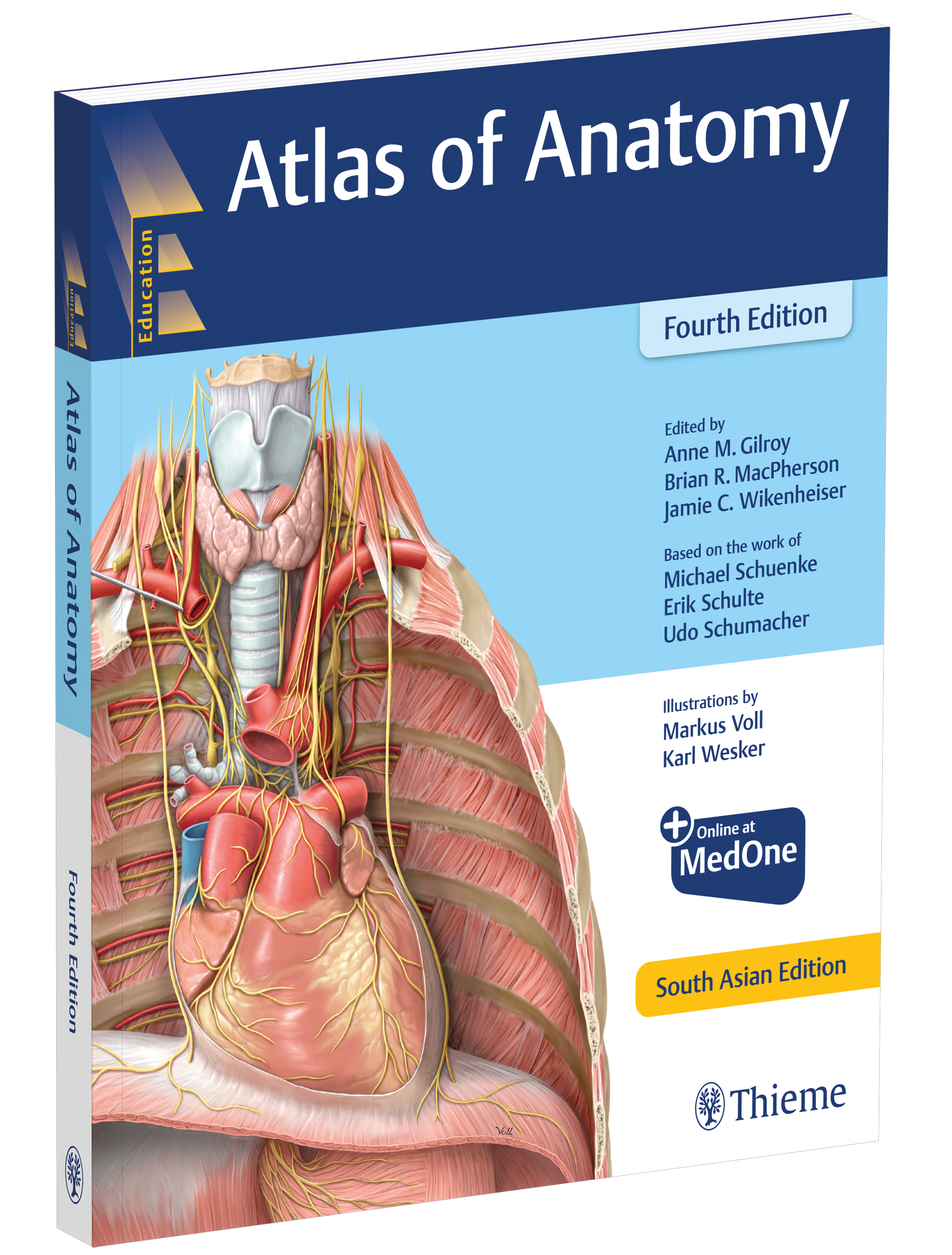
Atlas of Anatomy, Fourth Edition
Gilroy | MacPherson | Wikenheiser
Publication Date : 2021, Binding / Paperback | Edition / 4 | Pages / 780
ISBN : 9789390553464
NOW IN THE FOURTH EDITION
- NewExpanded Radiology sections include over 40 new radiographs, CTs and MRIs
- NewA more dissectional approach to the head and neck region places neck anatomy before that of the head – the way most students dissect.
- NewAdditional images and tables detail the challenging anatomy of the peritoneal cavity, inguinal region, and infratemporal and pterygopalatine fossae.
- NewAlmost 30 new clinical boxes focus on function, pathology, diagnostic techniques, anatomic variation, and more.
- NewMore comprehensive coverage clarifies the complexities of the ANS, including revised wiring schematics.
- NewMuscle Fact spreads provide origin, insertion, innervation, and action.
- NewAn innovative, user-friendly format: every topic covered in two side-by-side pages.
- NewOnline images with “labels-on and labels-off” capability are ideal for review and self-testing.
.jpg)
.jpg)
.jpg)
Our Other Notable Anatomy Books